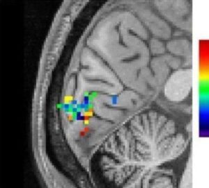
fMRI signals in the brain’s visual cortex as an individual views the checkerboard stimulus. The green, blue and dark blue spots represent signals produced within a fraction of a second after stimulation with dark blue being the fastest responding areas.
Credit: Lewis, et al. PNAS. Oct. 2016
Date: November 30, 2016
Source: National Institute of Biomedical Imaging and Bioengineering
Summary: Fast fMRI has been used to image rapidly fluctuating brain activity during human thought. fMRI measures changes in blood oxygenation, which were previously thought to be too slow to detect the subtle neuronal activity associated with higher order brain functions. The new discovery is a significant step towards realizing a central goal of neuroscience research: mapping the brain networks responsible for human cognitive functions such as perception, attention, and awareness.
https://www.sciencedaily.com/releases/2016/11/161130144008.htm
Recent Comments